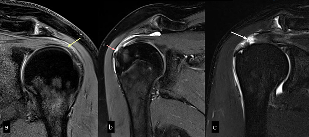
Typical magnetic resonance imaging scan showing the coracohumeral
Por um escritor misterioso
Last updated 23 outubro 2024


Shoulder MRI: normal anatomy

MAGNETIC RESONANCE IMAGING OF THE SHOULDER

Alteration of coracoacromial ligament thickness at the acromial undersurface in patients with rotator cuff tears - ScienceDirect

CT arthrogram of the shoulder joint: normal anatomy

Pain related to rotator cuff abnormalities: MRI findings without clinical significance - Bencardino - 2010 - Journal of Magnetic Resonance Imaging - Wiley Online Library

PDF] Radiological Variabilities in Subcoracoid Impingement: Coracoid Morphology, Coracohumeral Distance, Coracoglenoid Angle, and Coracohumeral Angle

Treatment of Isolated Subscapularis Tears - Cancer Therapy Advisor
Cureus, Role of Magnetic Resonance Imaging in the Evaluation of Rotator Cuff Tears

Magnetic resonance imaging of a male left shoulder showing narrowed

Narrowed coraco-humeral distance on MRI: Association with subscapularis tendon tear - ScienceDirect
Recomendado para você
-
 SCP-018 Superball, object class euclid in 202323 outubro 2024
 -
 scp-007 fan art (this one was challenging) : r/SCP23 outubro 2024
 -
 Pixilart - SCP-007-J by Mrdinosaur0223 outubro 2024
 -
 I love joke SCPs SCP Foundation Amino23 outubro 2024
 -
 LilyFlower's Workbench - SCP Foundation23 outubro 2024
 -
 Murakami's First Novels – Hear the Wind Sing / Pinball 1973 by Haruki Murakami Episode 007 : Infinite Gestation : Free Download, Borrow, and Streaming : Internet Archive23 outubro 2024
 -
 Create a SCPs featured in Confinement Tier List - TierMaker23 outubro 2024
 -
 Zera's SCPs23 outubro 2024
 -
 ArtStation - Source-engine Level Design23 outubro 2024
 -
SCP Foundation23 outubro 2024

você pode gostar
-
 Village Defense Tycoon codes (November 2023) - free gold and more23 outubro 2024
 -
 One Piece Film: Gold' heads back to theaters for anniversary23 outubro 2024
 -
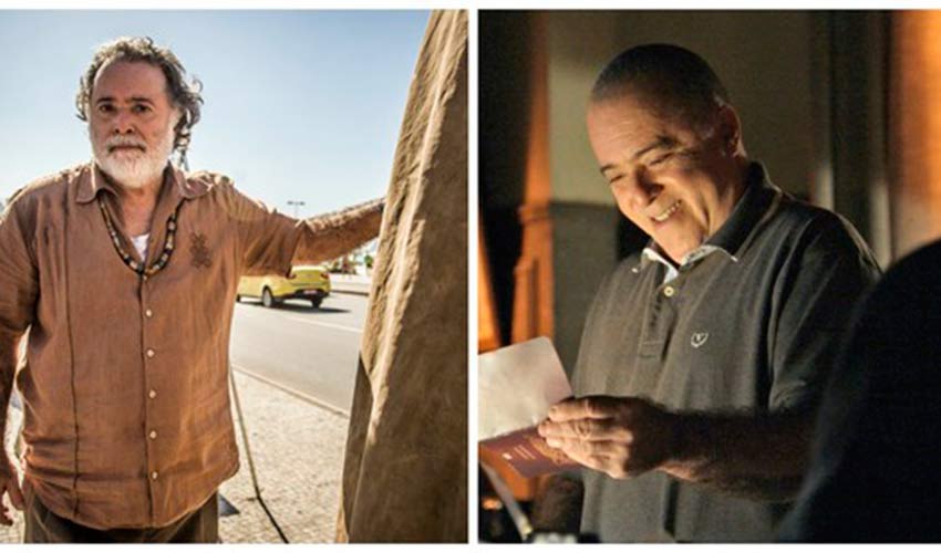 Tony Ramos muda visual em 'A Regra do Jogo'; veja transformações do ator - Entretenimento - Portal O Dia23 outubro 2024
 -
 Best Free Movie Streaming Sites in India in 2022 - Smartprix23 outubro 2024
 -
 TAG: Game Night Is It - Film Inquiry23 outubro 2024
 -
 Conjunto de vetores de ícones de porcentagem. ícones pretos de23 outubro 2024
 -
Subway Surfers Hypixel Forums23 outubro 2024
-
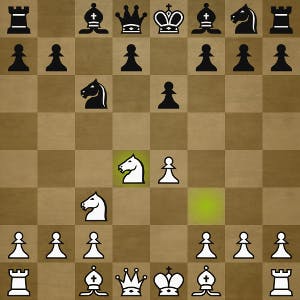 How to learn from your games at lichess23 outubro 2024
 -
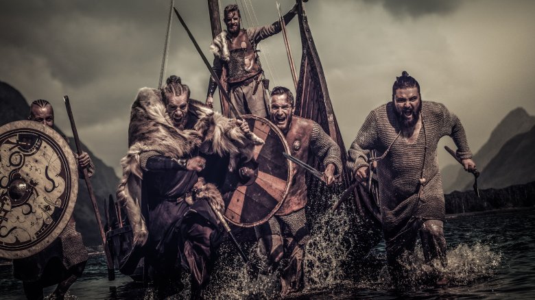 The Most Disturbing Thing About Viking Raids Isn't What You Think23 outubro 2024
 -
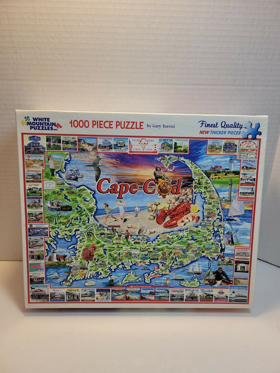 Quebra-cabeça White Mountain: Cape Cod MA. Quebra-cabeça de 100023 outubro 2024